About Us
Experienced Leaders in the Industry
The Vascular and Vein Center at Gulfcoast Surgeons has been one of the most respected vein clinics in Southwest Florida for over 30 years. Our surgeons, Dr. Abraham Sadighi, and Dr. Johan Escribano have performed thousands of vascular and vein surgeries with positive outcomes.
We focus on diseases of the vascular system that can range from harmless but unattractive spider veins to dangerous conditions such as peripheral artery disease. Our caring and dedicated team will help you identify problems and offer the best treatment options for you.
Varicose Vein Treatment
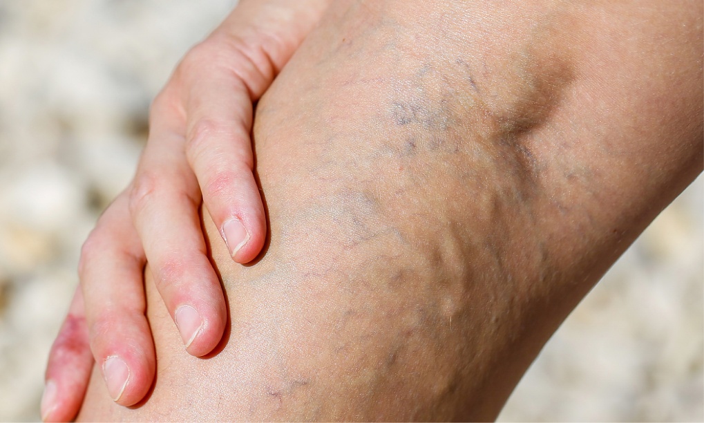
Varicose veins are enlarged, ropy, and often painful veins. They are usually blue or purple in appearance. Most people are familiar with varicose veins, though they might not recognize the name.
The word varicose means unusually swollen or knotty, which perfectly describes the veins snaking around the legs of many patients.
Our team provides expert treatment for varicose veins. We can help you love your legs again!

Vascular Surgery
Our vascular system is similar to a major transportation system throughout our body. Our veins and arteries supply our muscles and organs with blood and oxygen. Problems that arise within the vascular system can be critically important. Often symptoms of vascular diseases are non-existent or misinterpreted.
Our highly skilled vascular surgeons have years of experience fixing vascular issues. You’ll find our dedicated and caring staff to be a welcome comfort to you before, during, and after your surgery.
Spider Vein Treatment
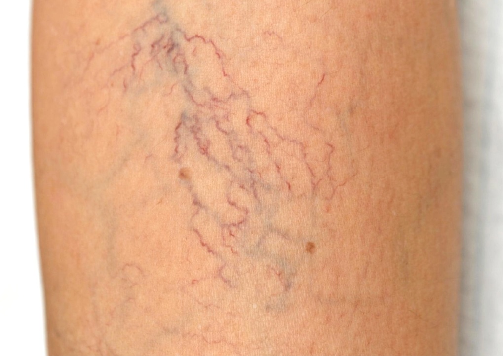
Spider veins are small red, purple, and blue “threadlike” veins are found on the surface of the skin. They occur on the legs, but can also be found on the face. Their only symptoms are cosmetic.
Spider veins are treated with sclerotherapy and cutaneous laser treatment. Our staff has years of experience treating spider veins and can help treat yours too!

Angio Suite
Patients who must undergo routine procedures such as angioplasty and stent typically have to go to a hospital outpatient facility, wait for hours, and pay exorbitant prices. Now, these procedures can be performed in our Angio Suite. We perform all our procedures in the peripheral vessels, such as the legs.
Through our state-of-the-art imaging machines, our surgeons can provide safe and effective procedures at a fraction of the cost and significantly quicker than a traditional hospital visit.
Our patients are extremely pleased with the convenience, cost savings, and caring treatment they receive from our staff.
Vascular Lab
Our state-of-the-art vascular lab allows our surgeons to conduct all necessary tests in-house allowing you to get the most comprehensive diagnosis and treatment options available.
These procedures are offered from our lab:
Ultrasound - Aortic
The abdominal aortic ultrasound is used to measure the aorta and detect or measure an aortic aneurysm. This procedure, like the other examinations, is painless. The ultrasound images allow physicians to determine if an aneurysm should be repaired or if it can be safely monitored without surgery.
Ultrasound - Mesentreric/Renal Artery
This ultrasound is used to detect a narrowing in the arteries that lead to the kidneys, and intestine, and is often done when patients are losing kidney function or have extremely high blood pressure, or have abdominal pain related to eating. This test also uses ultrasound waves, and typically takes two to three hours.
Patients should refrain from eating or drinking for eight hours prior to the test to allow the ultrasound waves to penetrate into the arteries.
Upper and Lower Arterial Study
An arterial evaluation is used to screen for peripheral arterial disease (PAD), or an arterial blockage in the legs. During this examination, the technologist will use a blood pressure cuff to measure the blood pressure in the legs. The pressure will be measured at the ankle, calf and thigh to screen for narrowing of the arteries.
Sometimes it is necessary for a patient to walk on a treadmill for five minutes while their blood pressure is being measured.
Venous Ultrasound
The venous Doppler examination is a study that is used to diagnose a blood clot, deep vein thrombosis (DVT), or venous insufficiency in the leg. Using ultrasound, a two-dimensional image is created enabling the doctor to see a blood clot and measure the size of the vein and see the functional status of the veins, especially in varicose vein patients.
Echocardiogram
An echocardiogram uses sound waves to produce an image of a beating heart. The image that is displayed allows the surgeon to see many of your heart’s structures and rhythm. Unlike an X-ray, the image of your heart moves in real-time instead of a static photo giving your doctor more information when evaluating cardiovascular conditions and helps with diagnosis and development of treatment options.
An echocardiogram is used to evaluate the function and anatomy of the heart, especially with respect to heart valves. It is used to evaluate and follow valvular heart disease and function of the heart.
Carotid Ultrasound
A carotid duplex examination uses ultrasound to study the carotid arteries, which are located in the neck. The test is used to detect a blockage or narrowing within the carotid arteries. A blockage of this type can cause a stroke, so detection may be lifesaving.
Dialysis Graft Evaluation
The dialysis graft evaluation examines access grafts in patients who are undergoing dialysis treatments. Since these grafts are accessed several times per week, they can develop scar tissue, which can in turn block blood flow and hinder the dialysis process. The ultrasound examination can detect the scar tissue and narrowing of the graft.
Bypass Graft Surveillance
Bypass graft surveillance is used to evaluate patients who have had vein bypass surgery in their lower extremities. The study examines whether the veins are functioning properly or if they have any narrowed areas. If a narrowing is found, the bypass vein can be repaired before the vein becomes entirely blocked.